Ultra High-Volume Scanners
PRODUCTS
-
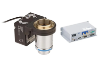
The P-725.xCDE1S PIFOC Scanner System offers a dynamic focus solution in the entry-level class, featuring travel ranges of 100 µm or 400 µm.
-
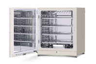
PHCbi brand's 8.1 cu.ft (230 L) Cell-IQ series CO2 incubator features a patented direct heat and air jacket system, dual IR sensor, SafeCell™ UV, and inCu-saFe® interior to help helps ensure precise temperature, CO2 levels, and contamination control for a precise and repeatable environment in a stackable, medium-sized capacity incubator for integrating with equipment and for scaling up cell culture production lines.
-
Accelerate your path to success with NetSuite by leveraging a top 3 NetSuite Partner in the world.
5 star, award-winning support in your corner
Optimize NetSuite for your industry, based on best practices, and get sound recommendations regarding processes and user scenarios based on years of expertise. We help our NetSuite clients and potential clients see what’s possible, set priorities, create a roadmap for accomplishment, and deliver results that matter.
When you choose Sikich, you choose a dedicated team of NetSuite consultants who spent years in a specific industry before they joined us, and who are committed to making a vital contribution to your business.
Get the connected, available and accountable support that your organization deserves by joining the ranks of our highly satisfied clients.
-
At ProPharma, we offer a unique approach to the traditional Clinical Research Organization (CRO) Full-Service Provider (FSP) model.
-
EirGenix provides client-oriented contract development and manufacturing services for biologics, especially monoclonal antibodies and biosimilars. Combining the capabilities of EirGenix’s strategic partner, Formosa Laboratories, Inc, a high potency API manufacturer, we offer integrated services for Antibody Drug Conjugate (ADC) development and manufacturing.
WHITE PAPERS AND CASE STUDIES
-
Prefabricated Viral Vector Modular BSL-2LS cGMP Facility In 13 Months
Learn about the modular BSL-2LS cGMP facility that was constructed to support 3 processing lines for German-based CDMO Vibalogics as the company sought to expand in the U.S. market for viral vectors.
-
The Savings Are Blowin' In At Port Washington Wastewater Treatment Plant
The municipal wastewater treatment plant in Port Washington, Wisconsin, is located on the picturesque shore of Lake Michigan, so it’s essential that their plant performs as designed.
-
Building Local Biomanufacturing Capacity In South Africa
Biovac evolved from a vaccine supplier to a biopharmaceutical innovator, providing a blueprint for expanding Africa's vaccine manufacturing capacity. Gain valuable insights from their journey.
-
Streamlining Study Start-Up For Accelerated Drug Development
What is the secret to safely expediting study start-ups? Explore our case study to find out how open dialogue, aligned expectations, and direct communication between Altasciences’ team leads and sponsor contributed to a clinical trial start-up of only 3.5 weeks.
-
Pioneering Cancer Research Meets Unparalleled Veeva Vault Integration
A biotech company revolutionizing cancer treatment faced delays due to a complex, error-prone document review process. Discover how integrating an innovative strategy transformed their workflow.
-
UV Treatment Upgrades In Wales
In order to improve the treatment performance (because of the continued cost to maintain compliance), and ensure that it would eventually have the treatment capacity to meet future population growth equivalent of up to 225,000, Swansea WwTW was in need of an equipment upgrade.
-
Revolution In The EU Pharmaceutical Legislation Ahead
Discover the impact revised pharmaceutical legislation will have on the industry by superseding regulations that have fostered the availability of safe and effective medicines for the past two decades.
-
How A Pharma Company Improved Yield By 1.5% In Just Three Months
A pharma company faced a 4% yield drop and variability at a manufacturing facility. Explore how they leveraged an AI-based platform to unify data, pinpoint inefficiencies, and enhance consistency in yields.
-
Revolutionizing Pharmaceutical Quality Control
Learn about a South Korean pharmaceutical company's QC laboratory and how the operation of its chromatography systems has been transformed by implementing a real-time monitoring system.
-
Advancing Multiomics Through Intelligent Automation
Modern automation tackles hidden pipetting errors and boosts reproducibility. Discover how your lab can scale confidently, reduce rework, and accelerate multiomic workflows with built-in traceability.
-
Enabling Digital Twins With Computational Fluid Dynamics Modeling
Embrace the transformative power of predictive modeling and digital twin technology to optimize bioprocess efficiency, ensure product quality, and drive innovation in biopharmaceutical manufacturing.
-
Streamlining Antibody Capture With Multi-Column Chromatography
Explore innovative solutions to overcome the limitations of traditional batch chromatography, improving efficiency, reducing costs, and optimizing production for complex biologics like monoclonal antibodies.
NEWS
-
Pharmaceutical Packaging Equipment Auction Announced6/20/2025
Don’t miss the latest Federal Equipment and Proxio Group auction featuring high-quality packaging equipment—pharma-origin with multi-industry use—including fillers, cappers, labelers, and more. The auction event runs June 23–July 9, 2025.
-
Premium Manufacturing, Packaging & Lab Equipment From Reckitt – Surplus Sale In Wilson, NC7/15/2025
Unlock major value for your facility with high-end, late model equipment from one of the most trusted names in health and hygiene. Federal Equipment Company, in partnership with Heritage Global Partners, is offering surplus process, packaging, and analytical lab equipment from a Reckitt facility in Wilson, North Carolina.
-
Samsung Introduces Galaxy XCover7 Pro And Galaxy Tab Active5 Pro: Ruggedized Devices For Frontline Excellence4/14/2025
Samsung Electronics today announced the new Galaxy XCover7 Pro and Galaxy Tab Active5 Pro, enterprise-ready devices designed to meet the demands of today’s fast-paced, high-intensity work environments.
-
Wagga Livestock Agents Lead The Way In Sheep And Goat eID Rollout5/14/2025
The Wagga Wagga Livestock Marketing Centre (LMC) has taken a major step forward in the transition to electronic identification (eID) for sheep and goats, successfully scanning nearly 15,000 individual electronic identification devices during last months record sale.
-
SmartVault Becomes First Document Management Platform To Integrate With Intuit ProConnect Tax1/28/2025
SmartVault, a leading provider of a cloud-based document management system (DMS) and client portal for accounting professionals, has expanded its ten-year strategic partnership with Intuit by integrating with ProConnect Tax, the industry-leading cloud-based tax software.

