High-Volume Scanners
PRODUCTS
-
Modern clinical trials require modern technology. Leverage the flexibility and speed of the cloud with TrialKit electronic data capture (EDC).
-
Protein A chromatography resin with excellent capacity and alkaline stability for cost-efficient and extremely robust mAb capture.
-
A successful clinical trial starts with an efficient protocol review.
With deep roots in academic research and clinical medicine, BRANY IRB is uniquely positioned to meet your cost and speed requirements. Get your clinical trial to the start line while ensuring participant health and safety.
-
At Applied StemCell, we specialize in producing high-quality differentiated cells from well-characterized normal iPSCs. Our cells offer the biorelevance of primary cells while eliminating the challenges associated with sourcing and variability across batches.
-
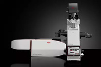
STELLARIS STED Microscope
Get faster to the truth
Our STED (stimulated emission depletion) technology joins the STELLARIS platform to provide you the fastest way of imaging beyond the diffraction limit. Obtain cutting-edge nanoscopy results in no time with astounding image quality and resolution, while protecting your sample. STED super-resolution allows you to study multiple dynamic events simultaneously, so you can investigate molecular relationships and mechanisms within the cellular context.
The seamless integration of STED and STELLARIS provides easy access to STED directly from the confocal interface, making it just a few clicks away. Now you can get more insights from your sample, because every detail matters.
WHITE PAPERS AND CASE STUDIES
-
How A Global Pharma Manufacturer Reduced Line Stops And Increased OEE
Gain insight into how a leading pharmaceutical company identified the root cause of long-standing inefficiencies, optimized performance, and productivity with unprecedented speed and accuracy.
-
Filtration System Rescues Bottled Spring Water Producer From Closure
Learn about the results a bottled water company witnessed from implementing the Aria FB-4 Water Filtration System to reduce their water filtration costs and achieve brand protection.
-
Mobile App And Wearable Integration Collects Longitudinal Data
Get an overview of SISCAPA's utilization of eTechnologies, specifically ePRO, and how their partnership with CDS accurately demonstrates the improvement of data collection and analysis in the biotechnology industry.
-
Advancing Multiomics Through Intelligent Automation
Modern automation tackles hidden pipetting errors and boosts reproducibility. Discover how your lab can scale confidently, reduce rework, and accelerate multiomic workflows with built-in traceability.
-
Simplify CAPA In 7 Steps
Discover how to streamline corrective action/preventive action (CAPA) management in regulatory environments in 7 steps.
-
Is Sustainability The Key To Agile Biopharma Manufacturing?
In biologics manufacturing, change is constant. Discover how new innovations and a focus on sustainability have gotten 62% of executives to prioritize eco-friendly practices, which reflects a commitment to long-term viability.
-
Leveraging High-Pressure Sterile Filtration For Highly Viscous Solutions
This research demonstrates the potential of high-pressure sterile filtration to enhance efficiency, reduce waste, and accelerate the development of innovative therapies.
-
Sulzer Mixers Designed To Tackle Digested Sludge
Discover how a wastewater treatment plant solved its mixing problem that was caused by the high sludge content.
-
Enhancing Treatment Capacity For Simmons Foods
Learn how Simmons Foods resolved their wastewater treatment issues by replacing their aeration equipment, resulting in increased treatment capacity, improved removal rates, and reduced operating costs and energy consumption.
-
A Synergy Of Excellence: Partnership During Unprecedented Times
Learn how a partner with the right experience and capabilities is crucial to support accelerated GMP manufacturing and ensure novel vaccines and therapeutics receive regulatory approval.
-
How To Manage Burgeoning Data Traffic On A Finite RF Spectrum
Explore strategies and innovations to unlock higher-frequency bandwidths, which are crucial for sustaining technological advancement and meeting the escalating demands of our connected future.
-
Buffalo, NY, Starts Successful Verification Program
Verifying service line materials can be a daunting undertaking, especially when the system is old and the staff is small. Read about how Buffalo Water addresses these challenges.
NEWS
-
CamScanner Launches Magic Pro Filter To Make Scanned Documents Perfect With Just One Click2/22/2024
CamScanner, a global leader in document scanning apps with over 300 million users, recently unveiled a revolutionary new tool enabling users to make their scanned documents perfectly clear with just one click. The cutting-edge "Magic Pro Filter" feature, powered by artificial intelligence, automatically detects blurs, and shadows in scans and effortlessly removes them.
-
FileCenter 12 Introduces Two-Factor Authentication And Dark Mode To Enhance Document Security For Small Businesses9/23/2025
FileCenter, the leading document management software for small and medium-sized businesses, today announced the release of FileCenter 12, featuring significant security enhancements, user experience improvements, and advanced document processing capabilities.
-
SmartVault Becomes First Document Management Platform To Integrate With Intuit ProConnect Tax1/28/2025
SmartVault, a leading provider of a cloud-based document management system (DMS) and client portal for accounting professionals, has expanded its ten-year strategic partnership with Intuit by integrating with ProConnect Tax, the industry-leading cloud-based tax software.
-
Transforming IDV Deployment: Regula Introduces Innovative Testing Service1/25/2024
Before adopting an IDV system, it’s imperative to conduct thorough testing, especially for large-scale businesses.
-
Field Squared Secures Innovative Technology Patent, Enhancing Efficiency And Customization For Field Service Automation8/1/2024
Field Squared, the industry-leading field service automation platform provider, is thrilled to announce the granting of a new patent for its cutting-edge system and method for constructing and displaying a variety of forms and documents through a client-side user interface.


