
New Optical Method Promises Faster, More Accurate Diagnosis Of Breast Cancer

Illinois researchers report on data-based method to replace manual inspection of tissue
A new optical method for more quickly and accurately determining whether breast tissue lesions are cancerous is described by University of Illinois researchers in the Journal of Biomedical Optics, published by SPIE, the international society for optics and photonics.
In "Breast cancer diagnosis using spatial light interference microscopy," published 20 August, University of Illinois at Urbana-Champaign and Chicago researchers Hassaan Majeed, Mikhail Kandel, Kevin Han, Zelun Luo, Virgilia Macias, Krishnarao Tangella, Andre Balla, andGabriel Popescu report on a quantitative method for diagnosing breast cancer using spatial light interference microscopy (SLIM).
Because this method is based on quantitative data rather than a subjective assessment, the researchers expect that these preliminary results show the potential of their technique to become the basis for an automated image analysis system that would provide a fast and accurate diagnostic method.
"Conventional methods for diagnosis of breast cancer have several limitations, including observer discrepancy," said YongKeun Park, a professor at KAIST and a guest editor of the special section on Quantitative Phase Imaging in Biomedicine in which the article appears.
When an abnormality in the breast is discovered, standard practice is for the physician to take a tissue biopsy, which is then stained to provide enough contrast for a pathologist to study key features of the tissue under a microscope. The tissue analysis is done manually. Due to variations such as staining intensity and the illumination used, the process does not lend itself to automation.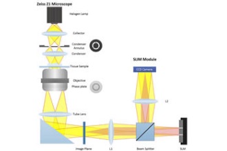
Manual inspections, however, are subject to investigator bias, and the process is time-consuming. This can, in some cases, result in late diagnosis -- a critical shortcoming given that early diagnosis significantly improves chances of survival.
Using the breast tissue biopsies of 400 different patients, researchers selected two parallel, adjacent sections from each biopsy. One was stained and the other left unstained.
The unstained samples were analyzed using a SLIM module attached to a commercial phase contrast microscope to generate interferograms -- photographic images derived from data based on how the tissue refracts light.
Four interferograms were used to produce one quantitative image showing areas with different refractive properties in different colors. The boundary between tumors and the cells around them were clearly delineated, making it possible to assess whether the tumors were malignant or benign.
More information about the technology and related applications can be found on the Quantitative Light Imaging Laboratory's website: www.light.ece.illinois.edu.
Breast cancer is the second most common form of cancer diagnosed worldwide, accounting for about 12% of all cancers diagnosed in 2012, according to the International Agency for Research on Cancer. Employing quantitative optical phase measurements, proposes an automated process that relies on quantitative imaging parameters for fast and accurate diagnosis.
Lihong Wang, Gene K. Beare Distinguished Professor of Biomedical Engineering at Washington University in St. Louis, is editor-in-chief of the Journal of Biomedical Optics. The journal is published in print and digitally in the SPIE Digital Library, which contains more than 430,000 articles from SPIE journals, proceedings, and books, with approximately 18,000 new research papers added each year.
About SPIE
SPIE is the international society for optics and photonics, an educational not-for-profit organization founded in 1955 to advance light-based science and technology. The Society serves nearly 264,000 constituents from approximately 166 countries, offering conferences and their published proceedings, continuing education, books, journals, and the SPIE Digital Library in support of interdisciplinary information exchange, professional networking, and patent precedent. SPIE provided more than $4M in support of education and outreach programs in 2014. For more information, visit www.spie.org.
Source: SPIE